Knee
Knee Anatomy
The knee is a complex joint made up of different structures including bones, tendons, ligaments and muscles. They all work together to maintain normal function and provide stability to the knee during movement.
Having a well-functioning healthy knee is essential for our mobility and ability to participate in various activities. Understanding the anatomy of the knee enhances your ability to discuss and choose the right treatment procedure for knee problems with your doctor.
Bones of the Knee
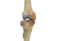
The knee is a hinge joint made up of two bones, the thighbone (femur) and the shinbone (tibia). There are two round knobs at the end of the femur called femoral condyles which articulate with the flat surface of the tibia called the tibial plateau. The tibia plateau on the inside of the leg is called the medial tibial plateau, and on the outside of the leg it is called the lateral tibial plateau.
The two femoral condyles form a groove on the front (anterior) side of the knee called the patellofemoral groove. A small bone called the patella sits in this groove and forms the kneecap. It acts as a shield and protects the knee joint from direct trauma.
A fourth bone called the fibula is the other bone of the lower leg. This forms a small joint with the tibia. This joint has very little movement and is not considered a part of the main joint of the knee.
Articular Cartilage and Menisci of the Knee
Movement of the bones causes friction between the articulating surfaces. To reduce this friction, all articulating surfaces involved in movement are covered with a white, shiny, slippery layer called articular cartilage. The articulating surface of the femoral condyles, tibial plateaus and the back of the patella are covered with this cartilage. The cartilage provides a smooth surface that facilitates easy movement.
To further reduce friction between the articulating surfaces of the bones, the knee joint is lined by a synovial membrane which produces a thick clear fluid called synovial fluid. This fluid lubricates and nourishes the cartilage and bones inside the joint capsule.
Within the knee joint, between the femur and tibia, there are two C shaped cartilaginous structures called menisci. Menisci function to provide stability to the knee by spreading the weight of the upper body across the whole surface of the tibial plateau. The menisci help in load- bearing by preventing the weight from concentrating onto a small area, which could damage the articular cartilage. The menisci also act as a cushion between the femur and tibia by absorbing the shock produced by activities such as walking, running and jumping.
Ligaments of the Knee
Ligaments are tough bands of tissue that connect one bone to another bone. The ligaments of the knee function to stabilize the knee joint. There are two important groups of ligaments that hold the bones of the knee joint together, collateral ligaments and the cruciate ligament.
Collateral ligaments are present on either side of the knee. They function to prevent the knee from moving too far during side to side motion. The collateral ligament on the inside is called the medial collateral ligament (MCL) and the collateral ligament on the outside is called the lateral collateral ligament (LCL).
Cruciate ligaments, present inside the knee joint, control the back-and-forth motion of the knee. The cruciate ligament in the front of the knee is called anterior cruciate ligament or ACL and the cruciate ligament in the back of the knee is called posterior cruciate ligament or PCL.
Muscles of the Knee
Muscles: There are two major muscles, the quadriceps and the hamstrings, which enable movement of the knee joint. The quadriceps muscles are in the front of the thigh. When the quadriceps muscles contract, the knee straightens. The hamstrings are in the back of the thigh. When the hamstring muscles contract, the knee bends.
Tendons of the Knee
Tendons are structures that attach muscles to the bone. The quadriceps muscles of the knee meet just above the patella and attach to it through a tendon called the quadriceps tendon. The patella further attaches to the tibia through a tendon called the patella tendon. The quadriceps muscle, quadriceps tendon and patellar tendon all work together to straighten the knee. Similarly, the hamstring muscles at the back of the leg are attached to the knee joint with the hamstring tendon.
Conditions
Knee Arthritis
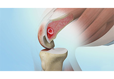
Arthritis is a general term covering numerous conditions where the joint surface or cartilage wears out. The joint surface is covered by a smooth articular surface that allows pain-free movement in the joint. This surface can wear out for several reasons; often the definite cause is not known. Arthritis often affects the knee joint.
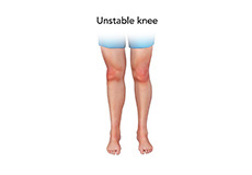
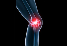
Unstable Knee
Unstable knee can be caused by sudden twisting of the knee, tears of the meniscus, ligament or capsule, osteoarthritis of the knee (wear and tear of the cushioning cartilage tissue between bones) and sports injuries.
.jpg)
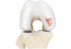
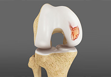
Chondral (Articular Cartilage Defects)
Articular cartilage does not have a direct blood supply to it, so has less capacity to repair itself. Once the cartilage is torn it will not heal easily and can lead to degeneration of the articular surface
Procedures
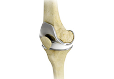
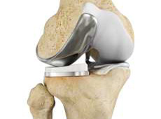
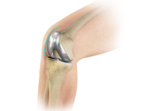
Total Knee Replacement
Total knee replacement, also called total knee arthroplasty, is a surgical procedure in which the worn out or damaged surfaces of the knee joint are removed and replaced with artificial parts.
Unicompartmental Knee Replacement
Unicompartmental knee replacement is a minimally invasive surgery in which only the damaged compartment of the knee is replaced with an implant. It is also called a partial knee replacement.
Revision Knee Replacement
Revision knee replacement surgery involves replacing part or all your previous knee prosthesis with a new prosthesis. Although total knee replacement surgery is successful, sometimes the procedure can fail due to various reasons and may require a second revision surgery.
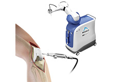
Robotic Assisted Knee Replacement
Robotic assisted knee replacement surgery is an alternative to the conventional knee replacement procedure. It is performed using robotic-arm technology that allows the surgeon to precisely perform the surgery through a smaller incision as compared to traditional surgery.
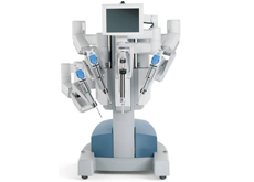
Robotic Assisted Partial Knee Surgery
Robotic assisted partial knee surgery is an innovative alternative to the conventional surgical procedure in patients suffering from degenerative knee diseases such as osteoarthritis. It is performed using robotic-arm technology that allows the surgeon to precisely perform the surgery through a smaller incision as compared to traditional surgery.
Outpatient Total Knee Replacement
Total knee replacement is the surgical treatment for knee arthritis, where the damaged knee is removed and replaced with an artificial knee implant. Traditionally performed as an inpatient procedure, total knee replacement surgery is now being conducted on an outpatient basis, allowing patients to go home the same day of the surgery.
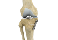
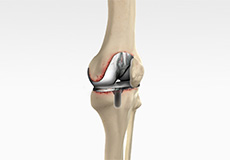
Knee Osteotomy
Knee osteotomy is a surgical procedure in which the upper shinbone (tibia) or lower thighbone (femur) is cut and realigned. It is usually performed in arthritic conditions affecting only one side of your knee and the aim is to take pressure off the damaged area and shift it to the other side of your knee with healthy cartilage.
High Tibial Osteotomy
High tibial osteotomy is a surgical procedure performed to relieve pressure on the damaged site of an arthritic knee joint. It is usually performed in arthritic conditions affecting only one side of your knee and the aim is to take pressure off the damaged area and shift it to the other side of your knee with healthy cartilage.
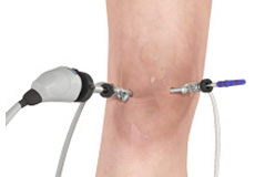
Knee Arthroscopy
Knee Arthroscopy is a common surgical procedure performed using an arthroscope, a viewing instrument, to diagnose or treat a knee problem. It is a relatively safe procedure and most of the patients are discharged from the hospital on the same day of surgery.
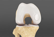
Cartilage Replacement
Cartilage replacement is a surgical procedure performed to replace the worn-out cartilage with new cartilage. It is usually performed to treat patients with small areas of cartilage damage usually caused by sports or traumatic injuries.
Cartilage Repair and Transplantation
Cartilage cell transplantation is a technique of transplanting healthy cartilage cells to replace damaged cartilage cells.
Autologous Chondrocyte Implantation
Autologous chondrocyte implantation (ACI) is a procedure to treat the articular cartilage defects of the knee. This procedure is effective for treating small areas of cartilage damage that causes pain and swelling and restricts the range of motion.
Subchondroplasty
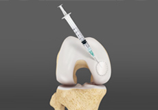
Subchondroplasty is a minimally invasive procedure that is performed to specifically repair chronic BMLs by filling them with a bone substitute material. The bone substitute is then slowly resorbed and replaced with healthy bone repairing the bone defect.

OATS
OATS is “osteochondral autograft transfer system”. It is one of the two types of cartilage transfer procedures and the other procedure is “Mosaicplasty”. Cartilage transfer procedures involve moving healthy cartilage from a non-weight-bearing area of the knee to a damaged area of the cartilage in the knee.